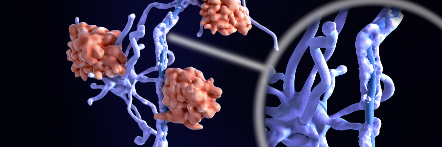
The underlying pathogenesis of MS remains an area of considerable interest and research and was one of the key themes examined during the 38th Congress of ECTRIMS [1]. Our understanding of the processes that govern lesion initiation, disease progression and remyelination remains limited, and it is possible that inflammatory changes seen early in the disease course could provide answers [2]. From the early stages of MS, the presence of pro-inflammatory and cytotoxic factors has been noted in the central nervous system (CNS) and shown to be associated with disease severity and clinical course.
Novel imaging techniques [3] and immune cell profiling [4] can both improve our understanding of the underlying pathogenesis of MS and provide novel insights that may facilitate the identification of new pharmacological targets, immunotherapies and personalised treatment approaches to prevent disease progression. It may also be possible to employ imaging techniques to more accurately monitor both demyelination and remyelination of MS lesions [5].
Inflammatory pathogenesis of MS
 MS has been described as a prototypically idiopathic neuroinflammatory disorder [6], with immune cell trafficking into the CNS considered a key component of its pathogenesis. Importantly, high-dimensional single cell characterisation of blood-borne immune cells present within the CNS of individuals with MS has revealed regional heterogeneity, with a signature distinct from that found in patients with other neurological diseases. By improving our understanding of the precise immune cells migrating into the CNS in MS, and the means by which this migration occurs, it may be possible to develop new diagnostic tools or treatment strategies.
It has also been postulated that the gut microbiome may play a central role in immune-mediated inflammatory diseases, with the microbiota influencing cell trafficking in the intestine [7]. Such hypotheses may align with observations of considerable aberrations in the gut microbiota of patients with MS [8] that have been found to be directly associated with blood biomarkers of inflammation, and provide further evidence in support of the role of the gut microbiome in inflammation, as discussed in a previous issue of ECTRIMS Insights [9].
Moreover, factors such as lipids and lipid mediators have been shown to play a major role in modulating CNS physiology and are essential to the regulation of neuro-inflammatory responses [10]. Research has demonstrated a disruption in the processing and efflux of lipids in foamy macrophages of individuals with MS, with lipid accumulation in phagocytes shown to induce an inflammatory lesion-promoting phenotype. Lipid metabolism can be a major determinant of CNS remyelination in individuals with MS, and so it is possible that therapeutic strategies which target the lipid-immune axis may promote lesion repair and encourage endogenous remyelination.
Promotion of remyelination to prevent neurodegeneration
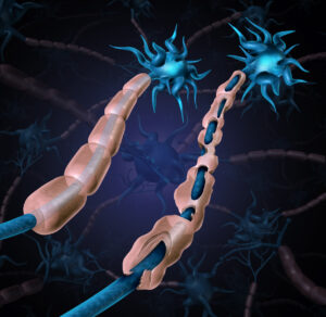
 Persistent demyelination is a key characteristic of MS, therefore strategies to encourage remyelination are required to prevent further neurodegeneration and assist recovery in individuals with MS [5]. Research presented at the 38th Congress of ECTRIMS described differences in the appearance of extracellular matrix surface structures in demyelinated lesions when compared with non-demyelinated areas. Moreover, the process of remyelination may be less effective in individuals with late-onset MS, possibly owing to the lower number of mature and active myelinated oligodendrocytes in normal and perilesional white matter; a finding also associated with higher levels of disability. The promotion of maturation of oligodendrocyte progenitor cells by neuregulin-1 has been proposed as a potential treatment strategy to stimulate remyelination.
Multiple animal models [11], including the toxin-induced demyelination cuprizone model [12], have been developed in an attempt to more fully elucidate the pathological mechanisms underlying demyelination in MS and to test potential therapeutic interventions. However, there are few research models that can reproduce the study of remyelination in the presence of ongoing inflammation, or that replicate the biological basis of chronic demyelination in MS. Nonetheless, despite the limitations of studying demyelination in toxin-induced animal models, such approaches may prove useful tools in identifying novel therapeutic strategies to promote CNS remyelination.
Promoting continued advances in research
ECTRIMS welcomes the continued research into the underlying pathogenesis of MS and applauds the efforts of the scientific community to constantly improve our understanding of how we might be able to halt disease progression and optimise patient care. We remain optimistic that ongoing research will continue to improve our knowledge of how best to treat individuals with all types of MS and provide a personalised approach to care.
***
ECTRIMS Insights articles are produced with an intent of being a neutral source of information sharing and objective analysis for the MS and neuroscience community. Unless otherwise stated, cited information in our articles does equivocate official endorsement from ECTRIMS.
***
REFERENCES
[1] ECTRIMS 2022. 38th Congress of the European Committee for Treatment and Research in Multiple Sclerosis. Available at: https://2022.ectrims-congress.eu/. Accessed January 2023.
[2] Scientific Session 12: The Earliest Events in MS. ECTRIMS 2022. Available at: https://vmx.m-anage.com/congrex/ectrims2022/en-GB/session/62797. Accessed January 2023.
[3] Hot Topic 4: New Ways of Imaging MS Pathology. ECTRIMS 2022. Available at: https://vmx.m-anage.com/congrex/ectrims2022/en-GB/session/62743. Accessed January 2023.
[4] Young Scientific Investigators’ Session 1: Immune Profiling. ECTRIMS 2022. Available at: https://vmx.m-anage.com/congrex/ectrims2022/en-GB/session/62757. Accessed January 2023.
[5] Scientific Session 3: Remyelination. ECTRIMS 2022. Available at: https://vmx.m-anage.com/congrex/ectrims2022/en-GB/session/62753. Accessed January 2023.